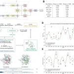
Understanding cell dynamics, regulation, and characteristics has been revolutionized by profiling tests at a previously unheard-of resolution. Nevertheless, the destructive nature of these techniques makes it difficult to monitor the temporal dynamics of living cells. Although it lacks genetic and molecular interpretability, Raman microscopy offers a unique way to report vibrational energy levels at subcellular spatial resolution. The researchers created Raman2RNA (R2R), an experimental and computational framework that uses multi-modal data integration, domain translation, and label-free hyperspectral Raman microscopy images to infer single-cell expression patterns in living cells. Raman images were used to link scRNA-seq profiles to paired spatial hyperspectral Raman images, and machine learning models were trained to infer expression profiles from Raman spectra at the single-cell level. In reprogramming mouse fibroblasts into induced pluripotent stem cells (iPSCs), R2R accurately inferred the expression patterns of numerous cell stages and fates, including MET cells, iPSCs, stromal cells, fibroblasts, and epithelial cells. This demonstrates how crucial spectroscopic content is to Raman microscopy.
Understanding of Methods Involved in Live Cell Tracking
The dynamic balance of extrinsic and internal programs determines the states and functions of cells. Numerous genes work together to coordinate the expression and function of these activities, which include cell proliferation, stress responses, differentiation, and reprogramming. These genes also interact with other cells and the environment to influence these processes.
Major advances in single-cell genomics and microscopy have enabled us to view gene programs and cell states at the genomic level, but these methods are destructive and involve tissue fixation, freezing, or cell lysis. Sophisticated computational techniques, such as velocity-based approaches and pseudo-time algorithms, can deduce dynamics from molecular profile pictures, but they still depend on assumptions that are difficult to test empirically. The movements of certain genes and programs within living cells can be observed by fluorescent reporters, although their use is constrained by the number of targets they can monitor, necessitates prior target selection, and frequently requires the use of genetically modified cells. The majority of dyes and reporters also need to be fixed, and they may also interfere with developing biochemical processes. Therefore, it is technically difficult to dynamically track the activity of several genes at once.
Raman Microscopy: Peering into Cellular Realms
Raman microscopy is a powerful analytical tool that uses the Raman scattering phenomenon. It provides a unique approach to tracking living cells and tissues since it provides chemical fingerprints of individual cells by reporting on the vibrational energy levels of molecules at a subcellular spatial resolution in a label-free and non-destructive manner. Raman microscopy may be used to characterize different cell types and states, discover germs resistant to antibiotics, diagnose pathological specimens like tumors non-destructively, and characterize the developmental stages of embryos, according to a groundbreaking study. However, the great dimensionality and complexity of the spectra, the spectral overlaps of biomolecules like proteins and nucleic acids, and the lack of unifying computational frameworks have hindered the breakdown of the underlying chemical profiles.
Raman2RNA: Bridging Raman Spectra to RNA Expression
Raman2RNA infers single-cell RNA expression profiles from label-free, non-destructive Raman hyperspectral pictures. Spatially resolved hyperspectral Raman pictures of living cells, selected marker smFISH data from the same cells, and scRNA-seq data from the same biological system are the inputs used by R2R. Next, R2R learns a model that connects spatially resolved hyperspectral Raman pictures to scRNA-seq using the smFISH data as an anchor. In the final phase, R2R employs this model to computationally derive the anchor smFISH measurements and single-cell expression profiles from hyperspectral Raman images. The outcome is a label-free live-cell inference of single-cell expression profiles.
Researchers created a high-throughput multi-modal spontaneous Raman microscope that allows for the automated acquisition of brightfield, fluorescence, and Raman spectra in order to streamline the data-collecting process. Specifically, the researchers used high-speed galvo mirrors and motorized stages to provide a large field of view (FOV) scanning and connected Raman microscopy optics to a fluorescence microscope where specific electronics automate measurements across different modalities.
The research showed that mouse-induced pluripotent stem cells (iPSCs) and mouse fibroblasts are two different cell types whose profiles can be inferred using R2R. This was accomplished by combining equal numbers of cells, plating them in a Petri dish, and performing live-cell Raman imaging. Fluorescence imaging was used for image registration and cell segmentation, and an Oct4-GFP, iPSC marker gene, was used. The Raman microscope’s excitation wavelength was selected to prevent phototoxicity and interference with cellular Raman spectra. To find marker genes for mouse iPSCs and fibroblasts, smFISH was carried out after Raman and fluorescence imaging. Polystyrene control bead pictures or reference points were used to register GFP, HCR, Raman, and nuclei stain images. A low-dimensional embedding of hyperspectral Raman data revealed that the Raman spectra discriminated the two cell populations in a way consistent with the expression of their respective reporters.
Key Findings
- By combining Raman hyperspectral images with scRNA-seq data through paired multi-modal data integration, smFISH measurements, and translation, researchers published R2R for inferring expression profiles at single-cell resolution from Raman spectra of live cells.
- High-accuracy single-cell expression profiles were deduced by the researchers using co-embeddings of individual profiles as well as averages within cell types.
- Researchers also demonstrated that brightfield z-stack predictions performed poorly, highlighting the value of Raman microscopy in expression profile prediction.
Limitations of Raman2RNA and Possible Solutions
- Single-cell Raman microscopy still has a low throughput. Approximately 6,000 cells were profiled in this pilot investigation. Scientists expect multiple orders of magnitude throughput increases using developing vibrational spectroscopy techniques, such as photothermal microscopy or stimulated Raman Scattering microscopy, to match the throughput of massively parallel single-cell genomics.
- Scientists can use advances in computational microscopy to infer high-resolution data from low-resolution data, such as compressed sensing, to further increase throughput. This is possible because molecular circuits and gene regulation are structured, with strong co-variation in gene expression profiles across cells.
- By using techniques like seqFISH, merFISH, STARmap, or ExSeq to identify more anchor genes, we can improve prediction accuracy and capture more single-cell variance. Furthermore, we may project additional modalities from Raman spectra, including scATAC-seq, using single-cell multi-omics.
- Given the similarities in the overall independent embedding of Raman and scRNA-seq profiles, we expect computational methods such as multidomain translation to allow mapping between Raman spectra and molecular profiles without measuring any anchoring in situ.
Conclusion
Raman2RNA is a label-free, non-destructive framework that integrates Raman hyperspectral pictures with scRNA-seq data through paired smFISH measurements, multi-modal data integration, and translation. This allows for the inference of expression profiles at single-cell resolution from Raman spectra of living cells. The researchers used both co-embeddings of individual profiles and averages within cell types to infer single-cell expression profiles with a high degree of accuracy. Researchers also demonstrated that brightfield z-stack predictions performed poorly, highlighting the value of Raman microscopy in predicting expression profiles. The combination of Raman microscopy and Raman2RNA seems promising in the future for transforming precision medicine, drug discovery, and other research fields.
Article Source: Reference Paper | Reference Article | Code for R2R is available at GitHub | Control software for the multimodal Raman microscope is available at https://github.com/kosekijkk/multimodal-raman-acq
Learn More:
Deotima is a consulting scientific content writing intern at CBIRT. Currently she's pursuing Master's in Bioinformatics at Maulana Abul Kalam Azad University of Technology. As an emerging scientific writer, she is eager to apply her expertise in making intricate scientific concepts comprehensible to individuals from diverse backgrounds. Deotima harbors a particular passion for Structural Bioinformatics and Molecular Dynamics.


