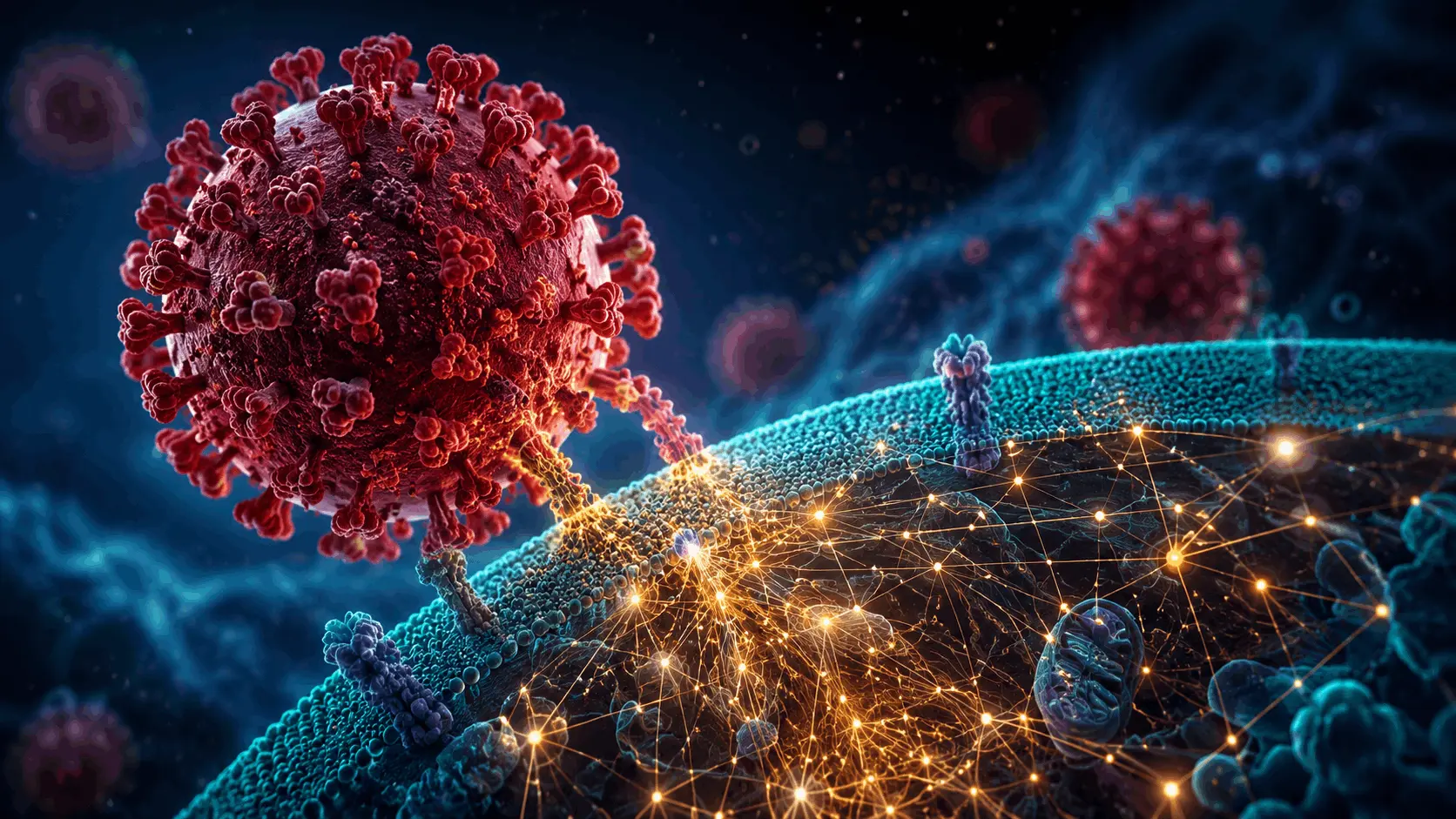
Every winter, the influenza A virus infects millions of people. In a handful of cells, it manages to do something very hard for scientists to observe: it enters a human cell, releases its own genetic material, and starts rewiring almost everything around it. We have known for decades that this rewiring happens, but seeing exactly which viral and human proteins touch each other, and where, inside a living, infected cell has been a genuine technical problem. A new study by researchers at EMBL Hamburg and their collaborators in Berlin has now managed to do exactly that and, along the way, has found a mechanism that nobody had described before.
Why was this hard to study
Most earlier work on how flu proteins interact with our own proteins relied on first breaking cells open. You lyse the cell, pull out the proteins you are interested in, and see what sticks to them. The problem is that once the cell’s internal walls and compartments are gone, proteins that would never have met inside a living cell can suddenly bump into each other in a test tube. Fragile, short-lived, or location-specific contacts are also easily lost in the process. So a lot of what we thought we knew about flu-host interactions may reflect what happens after the cell is destroyed, not what happens during infection.
To get around this, the team used a technique called in-cell cross-linking mass spectrometry, combined with a labeling method that flags newly synthesized viral proteins within infected cells. In simple terms, chemical cross-linkers were used to “freeze” proteins in place while they were still touching each other inside intact cells, and mass spectrometry was then used to read out which residues on which proteins had been locked together. This gave the researchers a snapshot of protein contacts as they actually existed during infection, not after the cell had been taken apart.
What they found
The dataset that came out of this work is large: thousands of cross-links covering hundreds of virus-to-host protein pairs, and nearly a thousand pairs involving human proteins interacting with each other in infected cells. Two results stood out.
The first involves haemagglutinin, the surface protein flu uses to latch onto and enter our cells. The team traced how this protein moves through the cell’s internal processing system, the endoplasmic reticulum and Golgi apparatus, picking up sugar modifications along the way. Several host proteins that help fold and process haemagglutinin were identified, including some that had never been linked to flu infection before. Turning these host proteins off, using RNA interference, reduced how well the virus could replicate, which is a good sign that the interactions the team mapped are functionally important and not just incidental contacts.
The second, and arguably more surprising, finding concerns tiny nuclear structures called paraspeckles. These are membraneless compartments inside the cell nucleus built around a long strand of non-coding RNA. The researchers found that infection consistently causes these structures to dissolve across every cell line and every flu strain they tested. This was not something they expected to see, and it occurred in a highly consistent way, suggesting it was not a side effect of a sick cell but a deliberate strategy on the virus’s part.
Digging further, the team found that two viral proteins, nucleoprotein and NS1, physically interact with the proteins that build paraspeckles. A viral enzyme called PA-X was also found to chew up the long RNA molecule that gives paraspeckles their structure. On top of that, the virus interferes with the host’s RNA polymerase II, the machinery that normally produces that structural RNA. So the disassembly of paraspeckles appears to be a three-pronged attack, involving direct protein interactions, RNA degradation, and interference with RNA production.
Why would a virus bother dismantling these structures? The likely answer is that paraspeckles normally hold onto RNA-binding proteins that the cell needs for other jobs, including parts of its antiviral response. When paraspeckles fall apart, those proteins are released, and the virus appears to redirect at least some of them to support its own replication.
Why this approach matters beyond flu
Perhaps the most useful part of this work is not any single interaction but the method itself. Because it captures contacts as they occur inside living, infected cells, this in-cell cross-linking approach avoids many of the artifacts that come from working with broken-open cells. The same combination of chemical cross-linking, mass spectrometry, and structural modeling with AlphaFold could, in principle, be applied to other viruses that hijack host cells in comparable ways, potentially including strains of pandemic concern such as H5N1.
For now, the study gives researchers a much more detailed and trustworthy map of how one of the most familiar human pathogens operates inside our cells, and a fresh set of host proteins worth closer scrutiny as potential targets for future antiviral drugs.
Article Source: Reference Paper | Reference Article
Disclaimer:
The research discussed in this article was conducted and published by the authors of the referenced paper. CBIRT has no involvement in the research itself. This article is intended solely to raise awareness about recent developments and does not claim authorship or endorsement of the research.
Follow Us!
Learn More:




Anchal is a consulting scientific writing intern at CBIRT with a passion for bioinformatics and its miracles. She is pursuing an MTech in Bioinformatics from Delhi Technological University, Delhi. Through engaging prose, she invites readers to explore the captivating world of bioinformatics, showcasing its groundbreaking contributions to understanding the mysteries of life. Besides science, she enjoys reading and painting.