
Scientists have developed a new CT technique using artificial intelligence (AI) to spot multiple myeloma-related bone diseases, using lower radiation doses than conventional CT, according to a study published in Radiology, a journal of the Radiological Society of North America.
After nearly two decades of development, the technology was used in clinical practice in 2021. Using the new technique, individual x-rays are directly converted into electric signals, which reduces detector pixel size and improves spatial resolution.
Multiple myeloma (MM)
Multiple myeloma (MM) is cancer that impacts white blood cells found in the bone marrow called plasma cells. Commonly appearing between the ages of 50 and 70, the disease affects both men and women equally.
In order to understand the potential for improved image quality in low-dose scans of the whole body better, Dr. Francis Baffour, the study’s lead author, a diagnostic radiologist at the Mayo Clinic in Rochester, MN, and colleagues studied the technology in people with multiple myeloma. There are approximately 80% of patients with multiple myeloma have lytic lesions, an area of bone destruction.
It is recommended that patients with associated bone disease undergo low-dose, whole-body CT scans to evaluate the disease and its treatment. In this setting, little is known about the photon-counting detector CT.
Photon-counting detector CT
CT is a medical imaging technology that implements X-rays to create cross-sectional images of internal organs. In photon-counting detector CT, the X-ray beam passes through the patient’s body and hits a scintillator material. The scintillator material emits photons at different wavelengths depending on their interaction with the X-ray beam. A photodiode detects these emitted photons and converts them into electrical signals. These electrical signals are then processed using a computer algorithm to produce a three-dimensional image of the patient’s anatomy.
According to Dr. Baffour, in addition, photon-counting CT provides ultrahigh-resolution images of large areas of the body with much better dose efficiency than standard CT.
The researchers performed low-dose, whole-body CT scans in 27 multiple myeloma patients with a median age of 68 years. They compared the images using both photon-counting detector CT and conventional low-dose, whole-body CT. Two radiologists evaluated the scans.
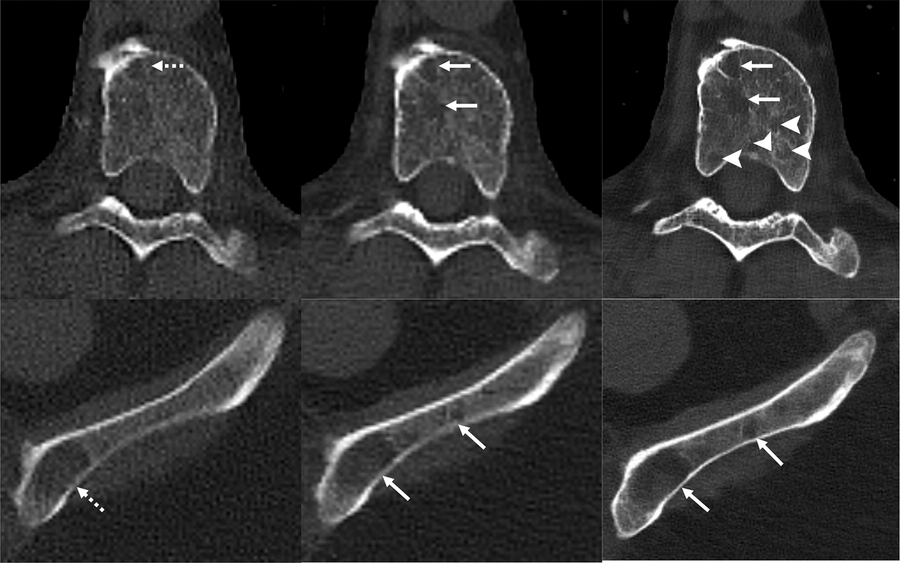
The Mayo Clinic CT Clinical Innovation Center’s CT clinical innovation team used deep learning AI to reduce noise in photon-counting CT images. The CT noise, or grainy appearance of cross-sectional images, is caused by an unwanted alteration in pixel values, which is often loosely defined as the noise in conventional CT. Photon-counting CT detectors with deep learning noise reduction produced sharper images and detected more tumors than conventional CT detectors. The team is excited that they were able to detect features of multiple myeloma disease activity more clearly using the photon-counting scanner.
We felt this was a prime example to showcase the ultra-high-resolution of photon-counting CT at low scanning doses.
Dr. Francis Baffour
The researchers are further planning to conduct follow-up studies on patients with multiple myeloma precursor states to see if photon-counting detector CT can spot bone lesions that would upstage them to active multiple myeloma.
Final thoughts
The researchers believe that this scanner could change the staging of diseases, influence therapy choices, and ultimately save lives. The researchers also want to examine photon-counting detector CT in other situations where low-dose protocols are beneficial, such as in pediatric or pregnant patients or screening procedures. The most important application is to reduce the radiation dose to patients during CT scanning. This will be possible with the development of photon-counting detectors with higher sensitivity and lower noise. Currently, several clinical research studies are underway to evaluate the use of photon-counting CT in various clinical settings. The results of these studies will be critical to the future of photon-counting CT in clinical care.
Story Sources: Reference Paper | Reference Article
Learn More:
Top Bioinformatics Books ↗
Learn more to get deeper insights into the field of bioinformatics.
Top Free Online Bioinformatics Courses ↗
Freely available courses to learn each and every aspect of bioinformatics.
Latest Bioinformatics Breakthroughs ↗
Stay updated with the latest discoveries in the field of bioinformatics.
Dr. Tamanna Anwar is a Scientist and Co-founder of the Centre of Bioinformatics Research and Technology (CBIRT). She is a passionate bioinformatics scientist and a visionary entrepreneur. Dr. Tamanna has worked as a Young Scientist at Jawaharlal Nehru University, New Delhi. She has also worked as a Postdoctoral Fellow at the University of Saskatchewan, Canada. She has several scientific research publications in high-impact research journals. Her latest endeavor is the development of a platform that acts as a one-stop solution for all bioinformatics related information as well as developing a bioinformatics news portal to report cutting-edge bioinformatics breakthroughs.


 to spot multiple myeloma-related bone diseases.)