Scientists from the Chinese Academy of Sciences, China, introduce SONAR, a novel model for cell-type deconvolution from spatial transcriptomics data. This method, termed Spatially weighted pOissoN-gAmma Regression (SONAR), utilizes a geographically weighted regression framework that leverages neighboring information to refine the local estimation of regional cell type composition. By incorporating an elastic weighting step, SONAR effectively filters out dissimilar neighbors, mitigating the risk of introducing unrealistic biases. Additionally, through preliminary clustering steps, SONAR excels in directly modeling the raw counts present in spatial transcriptomic data. The remarkable performance exhibited by SONAR sparks enthusiasm for its potential to harness the wealth of information within spatial transcriptomics resources.
Ameliorating Cell-Type Deconvolution Strategy of Spatial Transcriptomics Data
The researchers address the limitations of previous approaches to Spatial Transcriptomics (ST)-generated spatial information deconvolution algorithms. The previously developed deconvolution algorithms addressed the major issue of inferring a probable faulty measurement of RNA transcripts in spatial locations or spots due to the poor spatial resolution of ST data.
The authors articulate that although the concept of closely organized cells in a tissue tends to show similar features, it shouldn’t be generalizable for histological sections consisting of heterogenous cells, especially at the boundaries where cells of different functions can be closely situated. Without considering this ‘edge effect,’ an inherent bias would be propagated while demarcating cell-type proportions by those previous models. The spatially weighted pOissoN-gAmma Regression model, SONAR, aspires to rectify these gaps for cell-type deconvolution with spatial transcriptomic data.
Spatial Transcriptomics Over Conventional Transcriptomics
The study of entire gene transcripts, or RNA synthesized by the transcription machinery at a certain timeframe under the influence of particular circumstances, is called transcriptomics. For decades, high-throughput technologies mediating the acquisition of transcriptomics or the quantification of gene expression levels focusing on a single cell have been the backbone of many important and intriguing researches.
However, taking into account the intricate, highly specialized organ systems and respective tissue systems in organisms, it is obvious and crucial to gather transcriptomics information of multiple heterogenous cells of the tissue and to cognize the relative locations of cells in the histological section in order to integrate the previously neglected cell’s spatial contexts with an aim to acquire a comprehensive roadmap of the cell biology, tissue architecture, and gene expression patterns.
This novel breakthrough approach,’ Spatial Transcriptomics,’ presents scientists to explore a spatially resolved, high-dimensional assessment of gene activity executed in the tissue section and locate the part of the tissue where the activity is occurring. Spatial Transcriptomics puts emphasis on the relative positional contexts of cells with respect to their neighboring cells and other non-cellular structures, essentially because the microenvironment of the cell and inter-cellular signaling actively dictate several downstream gene expression pathways and brings variety at the transcriptome level of the same tissue.
Characterizing the spatial distribution of mRNA molecules facilitates an incredible scope to unravel cellular and subcellular architecture, cellular heterogeneity, cell states and functions of the sample, and most significantly, is useful in ameliorating perspectives regarding biological phenomena in healthy and diseased conditions that can’t be obtained by applying conventional techniques. In spatial transcriptomic data (ST data), the resolved relative spatial localization of spots provides valuable information about neighboring contexts.
SONAR Model: Architecture and Features
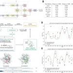
SONAR models the raw counts in ST data directly with a Poisson-Gamma mixture distribution, which is able to account for the overdispersion effect. Then, it applies a geographically weighted regression framework that adopts a kernel regression methodology to fully utilize the spatial information in order to decipher cell-type composition at each spatial spot by borrowing information from neighbors.
Notably, SONAR follows a pre-clustering step with the Louvain algorithm to avoid bias arising due to edge effects and also comprises an elastic weighting strategy to adaptively adjust the spatial kernel weights according to expression similarity. Consequently, dissimilar neighboring spots that are unlikely to share a common cell type composition are effectively screened, preventing the erroneous introduction of local estimation bias in transition regions with sharp boundaries.
Without that step, SONAR’s performance in Jump-transition between subregions is found to be low and exhibits sharp errors at the border of two subregions. SONAR outputs the spatial map of cell types and gives the detailed composition of each spot. Moreover, the evaluation investigations suggest the SONAR is robust enough to handle variation in cell composition, delivers improved characterization of regional co-localization of cell types, and consistently exhibits its performance with the lowest estimated error compared to other models.
Remarkably, even in real tissue samples, including the mouse brain, the human heart, tumors related to liver cancer, and human pancreatic ductal adenocarcinoma, it is capable of capturing the spatial structure of cell composition. SONAR can elucidate the detailed distributions and co-localization dynamics of immune cells around tumor leading edge regions. In addition, the cell type compositions determined by SONAR correspond to the clinical status of patients.
Conclusion
Optimization of ST technologies holds prospects for extracting novel, meaningful information and can upgrade our current understanding of biological processes and pathogenesis. Harboring ST data is challenging due to limitations in acquiring effective resolution of spatial information and further deconvolution of the sophisticated data. In this context, SONAR maps region-specific cell types that are extremely important for attaining a comprehensive landscape of heterogenous cells in the tissue and helpful in understanding spatial functional distribution. SONAR effectively omits the propensity to gather biases while investigating tissues with complex spatial patterns. The evaluation studies demonstrate SONAR’s capability to unravel important aspects of ST data.
Article Source: Reference Paper | The R package of SONAR is available on GitHub
Learn More:
Aditi is a consulting scientific writing intern at CBIRT, specializing in explaining interdisciplinary and intricate topics. As a student pursuing an Integrated PG in Biotechnology, she is driven by a deep passion for experiencing multidisciplinary research fields. Aditi is particularly fond of the dynamism, potential, and integrative facets of her major. Through her articles, she aspires to decipher and articulate current studies and innovations in the Bioinformatics domain, aiming to captivate the minds and hearts of readers with her insightful perspectives.


