
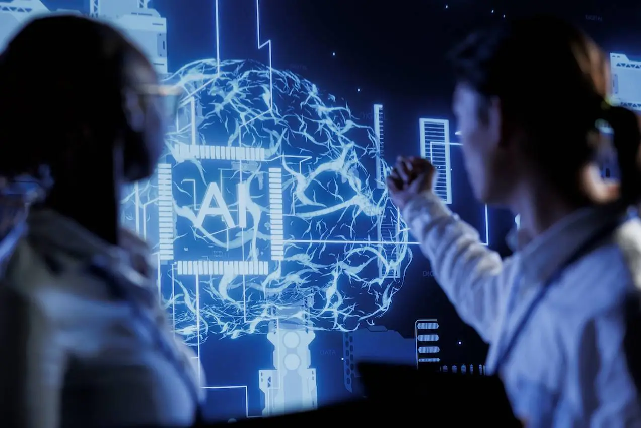
Artificial intelligence (AI) has become widely accessible, transforming from a specialized resource into a common tool for cancer researchers. It is beneficial for every cancer researcher to understand these tools. In this article, we’ll explore how AI is revolutionizing cancer research as well as some resources reported recently in the Nature Reviews Cancer journal by the scientists at Technical University Dresden, Germany, that could aid non-computational cancer researchers in their quest to save lives.
Consider an onco-pathologist, spending hours reviewing biopsies from tumor tissues for diagnosis.
He looks at his daughter, scribbling in her notebook.
“Weren’t you going to review your essay?”
“Yeah, I finished it.”
He was taken aback. “How?”
“Put it through GPT. It helped me summarize my mistakes. Now let me do math.”
He was impressed, wishing he could do the same.
Fortunately, a myriad of AI-based solutions remain unexplored, providing an opportunity to streamline cancer research endeavors.
Rise of Artificial Intelligence
Since the advent of this concept, artificial intelligence has been true to its name- artificial. It was mainly theoretical and lacked real-world practical applications. Recently, AI has achieved a human-level performance, especially in biomedical research. Hence, cancer researchers must have a certain level of AI literacy to leverage its full potential.
Artificial Neural Networks
Many of the present-day AI applications depend on ANNs. What are they?
Artificial neural networks (ANNs) are based on the most complex computer in the world- our brain. Similar to the structure of our brain, a neural network consists of interlinked units called neurons. There are typically three types of layers:
- The input layer receives data.
- The output layer produces results.
- Hidden layers perform most computations.
Deep Neural Networks and Deep Learning
A neural network with many hidden layers is called a deep neural network (deep learning). Deep networks can learn complex patterns by building higher levels of understanding and representation from data. In cancer research today, AI and deep learning are often used interchangeably, focusing on training networks with large datasets to adjust parameters and minimize output errors. Machine learning is a subset of AI that enables computers to learn from data and make predictions using statistical methods. Deep Learning is further a subset of ML that employs neural networks for hierarchical representation of data.
Branches of Machine Learning
Supervised Learning:
A model is trained over training data consisting of inputs and the ground-truth labels and is tested on unseen data. The model learns patterns present in the training data to make accurate predictions on the test set.
Unsupervised Learning:
No training data is available. The model is made to learn patterns themselves. It is commonly used in clustering, anomaly detection, and association tasks.
Reinforcement Learning:
This learning combines the system/model and its environment. The system takes certain actions based on rewards and feedback from the environment. The goal is for the system to learn a policy that maximizes the cumulative reward over time. Now that we have understood the basics let us dive into the applications of such models in cancer research.
Applications of AI in Cancer Research
AI in Biomedical Image Analysis
Automating the task of image analysis (Computer Vision) was a great boon for this industry. Breakthroughs in deep learning in the 2010s, like CNNs and large datasets, made computer-based image analysis more versatile for complex tasks, improving cell detection in microscopy images and enabling applications like cancer prognosis based on lymphocyte enumeration using tools like StarDist.
Cellular and Molecular Imaging Analysis
Deep learning can classify cells as “dead” or “alive” by detecting them in phase-contrast microscopy. Commercial platforms like the Incucyte AI Cell Health Analysis Software Module by Sartorius AG use deep learning for cell analysis.
Tools Available:
- QuPath is a software for viewing gigapixel microscopy images, enabling access to external ML models like StarDist. No knowledge of programming is required.
- ImageJ, ImageJ2, and Fiji are the standard tools for image viewing and analysis tasks in biology, including viewing multichannel, multidimensional images.
Computational Pathology
Tasks such as predicting genetic alterations, the mutation status of individual genes, defective DNA repair mechanisms, or tumor mutation burden from hematoxylin and eosin (H&E)-stained tissue from pathology slides are easily done using deep learning.
Tools Available:
- FastPathology, TIAToolbox, CLAM, and STAMP are built on top of general-purpose data processing and machine learning frameworks, such as PyTorch, MONAI, OpenSlide, and LibVips. Knowledge of Python and image processing is required.
Radiology
Radiology methods like CT, MRI, and PET are used in cancer detection and monitoring. Machine learning models depend on generated radiomics features, whereas deep learning models determine the features themselves, taking only the raw data as input.
Tools Available:
- Pyradiomics: A tool to extract radiomics features from region-of-interest in medical images.
- Aview LCS, syngo.CT Lung CAD, Samsung Auto Lung Nodule Detection, and InferRead CD Lung: These are medical devices that improve the detection of lung cancer in radiology.
We’ve discussed image analysis a lot, but what about words? Words and sentences can be used as extensive inputs for ML and DL models to produce accurate results. Let us look into their functioning.
LLMs in Cancer Research
Natural Language Processing is the analysis of human languages using computers. Large Language Models are based on transformer architecture and generative AI models and are used for NLP tasks. LLMs can analyze, translate, extract information from medical reports, and provide medical recommendations. In cancer research, they offer various benefits like idea refinement, skill acquisition, and efficient dissemination of findings.
OpenAI’s ChatGPT uses Self-Supervised Learning and is a foundation model. Foundation models are DL models that are pre-trained on a diverse dataset using SSL and can be trained on various data types, including images, text, or a combination of both.
Applications of LLMs in Different Tasks:
Zero-Shot Applications:
LLMs are used directly in a task without prior specific training. It depends greatly on the efficiency of prompt engineering. For example, You ask GPT to generate an essay on “Cancer Research.”
Beyond Zero-Shot:
LLMs are provided with some examples of the expected output. It depends on in-context learning. For example: You ask GPT to generate an essay on “Cancer Research” and also provide examples of some required subheadings like: “Tumor Image Analysis,” “Using ImageJ,” etc.
Retrieval-Augmented Generation:
LLMs create a stacked vector of documents to refer to whenever needed. For example:- Along with the prompt, you also pass research papers for GPT to use later.
Tools available:
- GPT-4, Gemini-1.5, Claude 3, Llama, and Mixtral are some LLMs available on the web.
We have discussed the various ways AI can be used for cancer research. Let’s finally look into the latest advancements and innovative uses of artificial intelligence in this field!
New Frontiers in AI Applications
Multimodal AI:
Integrating diverse data types, such as images and clinical patient information, into deep learning models has enhanced accuracy in cancer research.
Vision LLMs:
These do not require fine-tuning as they are trained on large datasets with images and text, which help them process and generate both modalities simultaneously. Llava-Med and PathChat can be used in biomedical analysis.
Drug Discovery:
Machine learning, especially deep learning models like AlphaFold, revolutionizes drug discovery by predicting protein structures and drug-protein interactions and generating new compounds.
Conclusion
AI can help cancer research and personalized medicine by developing more accurate biomarkers, identifying new drug targets, and simplifying drug development through hidden pattern recognition in molecular profiling. Cancer researchers should familiarize themselves with AI tools to navigate a future where AI is widely used in healthcare for better patient outcomes.
Article Source: Reference Paper Abstract
Follow Us!
Learn More:


Neermita Bhattacharya is a consulting Scientific Content Writing Intern at CBIRT. She is pursuing B.Tech in computer science from IIT Jodhpur. She has a niche interest in the amalgamation of biological concepts and computer science and wishes to pursue higher studies in related fields. She has quite a bunch of hobbies- swimming, dancing ballet, playing the violin, guitar, ukulele, singing, drawing and painting, reading novels, playing indie videogames and writing short stories. She is excited to delve deeper into the fields of bioinformatics, genetics and computational biology and possibly help the world through research!


