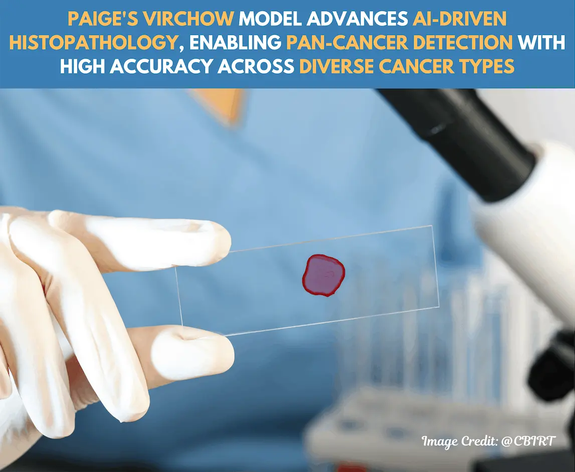
Computational pathology is the evolution of cancer diagnosis into a new era, with the application of artificial intelligence to understand complex histopathology images. Generally, pathologists spend a lot of time diagnosing based on H&E-stained slides. To reduce the efforts required, researchers from Microsoft Research, Memorial Sloan Kettering Cancer Center, NSW Health Pathology, St George Hospital, and the University of Rochester explain one of the latest developments of “Virchow” basis models that mark a huge leap forward in the field of computational pathology. Trained on huge amounts of digitized pathology images, it generally carries pretty robust capabilities to distinguish diverse variants of cancer, including rare types, with great precision in the direction of aiding clinical-grade diagnostics.
Role of the Foundation Models in Computational Pathology
Foundation models have been trained on vast amounts of data to produce generalizable representations suitable for numerous tasks without requiring large amounts of task-specific data. In the case of Virchow, this was done with over 1.5 million digitized histopathology slides. This level of scale alone is sufficient to allow the foundation model to detect subtle patterns across many tissue types and states of pathology-from benign lesions to highly complex malignant transformations. It is named after Rudolf Virchow, one of the precursors of cellular pathology, as many would know. It links extensive training data with an architecture that mirrors the ViT-H (Vision Transformer-High capacity) and uses 632 million parameters!
Virchow’s Applications: Pan-Cancer Detection and Rare Cancer Identification
The pan-cancer-detecting capabilities of Virchow are what differentiate this model. This capability allows the model to detect a wide range of cancer types rather than those specified origins in tissue. The model is trained on an extensive dataset of nine common and seven rare cancer types from Memorial Sloan Kettering Cancer Center (MSKCC). For evaluations, Virchow has shown an excellent AUC score in these cancers at 0.95, which is a notable task in pathology because it catches nuances that may indicate rare cancers.
Rare cancer detection is highly challenging because few samples are available, and rare cancer cell structures are difficult to differentiate from benign or common malignant patterns. Virchow outperformed or matched existing models in tests over detecting rare cancers, reaching an AUC of 0.937 in these cases. The ability of Virchow on single rare cancers was poor; cervical and bone cancers were undetectable, further suggesting improvements in the identification of cancers where the representation is less significant in the training dataset.
Improving the Detection of Biomarkers by Virchow
Virchow demonstrates an innovative capability to predict the biomarkers directly from H&E-stained tissue slides and has the potential to avoid second-line testing with IHC or molecular sequencing. This might accelerate the time towards diagnosis and reduce the distress of the patient since traditional detection of biomarkers often necessitates the repetition of a process multiple times. Within this study, the model can predict crucial biomarkers of breast, lung, and prostate cancers that are important in the personalization of cancer treatment.
Generalization and Robustness Across Diverse Clinical Settings
Virchow has proved robust on out-of-distribution (OOD) data, which basically means it holds accuracy on the data of other institutions and on tissue types that were not used in training. The clinical use calls for these requirements – the model actually sees patient data that is unlike those within a training set. For example, around 20% of the tissues in the test set of Virchow were never seen at train time, but the model remained consistent and gave high sensitivity and specificity.
Comparison to Clinical-Grade Models
Compared to clinical-grade AI products, which were specifically intended for the detection of prostate, breast, and lymph node cancer, the performance of the clinical models was better in those fields. However, the foundation model also turned out to be almost equivalent, with an AUC value of 0.980 for prostate cancer, 0.985 for breast cancer, and 0.971 to detect lymph node metastasis. This is an intriguing aspect because Virchow exceeded the models of specialists for some rare cancer forms, and there are some indications that it can be useful to applications in contexts where the detection of the totality of cancer is desired rather than task-specific specialization.
Methodology and Technical Advancements
Using the DINOv2 self-supervised learning algorithm, Virchow learns features using multiview training strategies from both local and global tissue regions. Virchow thus processes large WSIs as tiles of 224 x 224 pixels and produces embeddings that capture the complexity of tissue features. This produces slide-level outcomes for tasks like cancer type identification, biomarker prediction, and subtype classification synthesized by the aggregator model.
This multi-tasking ability and tile-based approach facilitate analyzing and learning on this highly diverse dataset, consisting of tissue slides prepared with diverse methods and cancer types; thus, the model can generalize over a wide range of histopathology images.
Challenges and Future Directions
Although it depicts a very high benchmark, Virchow has brought forward several challenges and future directions for the models of computational pathology. One of the ongoing challenges is the diversity in training data since histopathology data inherently contains a large number of morphologies and staining variations. Virchow’s success indicates the possibility that larger and more diversified datasets may improve the generalization of the model even further so that rare cancers can be detected. Furthermore, improvements in the model’s capability to make predictions at the slide level may optimize the diagnostic efficiency of the model by reducing the dependency bias of analysis at the tile level, thus further optimizing pathology workflow.
The efficiency of the model needs further enhancement. Virchow’s size might be large enough that it is not deployable in a real-time clinical setting due to high computational requirements, which are needed to achieve maximal accuracy. Model distillation, a technique that creates smaller and faster versions of large models, could help overcome this limitation and use foundation models in different clinical settings.
Conclusion
Briefly stated, this Virchow Foundation model represents an important step toward achieving AI in clinical pathology: It allows for a totally automatic cancer diagnosis encompassing cancers that are extremely rarely diagnosed and robustly predicts biomarkers directly from standard H&E slides. This helps minimize the efforts of doctors by quite a lot. Its size and performance thus demonstrate the real feasibility of foundation models in pathology and open possibilities for tools that could dramatically simplify diagnostics!
Article Source: Reference Paper | Reference Article | The model can be accessed at Hugging Face | A public software development kit for leveraging foundation model embeddings to develop downstream WSI applications is available on GitHub.
Disclaimer:
The research discussed in this article was conducted and published by the authors of the referenced paper. CBIRT has no involvement in the research itself. This article is intended solely to raise awareness about recent developments and does not claim authorship or endorsement of the research.
Follow Us!
Learn More:
Neermita Bhattacharya is a consulting Scientific Content Writing Intern at CBIRT. She is pursuing B.Tech in computer science from IIT Jodhpur. She has a niche interest in the amalgamation of biological concepts and computer science and wishes to pursue higher studies in related fields. She has quite a bunch of hobbies- swimming, dancing ballet, playing the violin, guitar, ukulele, singing, drawing and painting, reading novels, playing indie videogames and writing short stories. She is excited to delve deeper into the fields of bioinformatics, genetics and computational biology and possibly help the world through research!

